ANATOMY OF THE EAR - MICROTIA & ATRESIA

OUTER EAR - Pinna Structure and Ear Canal
The Outer Ear consists of the Pinna or external ear and the ear canal all the way up to the Tympanic membrane or eardrum. The most important function of the pinna in hearing is the capture and localization of the sound.
MIDDLE EAR - Ear Bones and Atresia-Related Hearing Loss
The middle ear is an air filled space or cavity about 1.3cm (one-half inch) across. The middle ear space houses three little bones called the malleus, incus and stapes. These bones conduct sound from the eardrum to the inner ear. You can see to the left how small our middle ear bones are. The bone pictured to the left is called the “stapes” and is actually one of the smallest bones in our body.
INNER EAR - Cochlea Function and Balance Explained
The inner ear consists of two structures: 1) The cochlea or hearing organ is shaped like a snail. The cochlea is fluid-filled and is lined by very tiny hair-like structures. When sound causes the eardrum and middle ear bones to vibrate, the last bone (stapes) moves in and out like the piston of a car. This causes a wave of fluid to move across the inner ear. This wave of fluid stimulates the hair-like structures. Through electrical impulses, the hair-like structures communicate with the hearing nerve that in turn communicates with our brain telling us that there is sound. 2) The semicircular canals help us sense our orientation and balance.
FREQUENTLY ASKED QUESTIONS
How does sound normally travel to the inner ear?
Sound is captured and localized by the pinna or external ear. This sound is then concentrated and directed into the ear canal until it strikes the eardrum. The eardrum then vibrates, causing vibration of the three middle ear bones. The last bone, called the stapes, then moves in and out into the inner ear. This causes a wave of fluid within the inner ear to stimulate the inner ear hair cells. These hair cells communicate with the hearing nerve via electrical impulses. The impulses from the hearing nerve tell our brain that we are hearing sound.
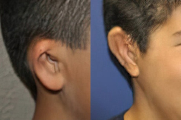
How can a microtia patient hear without an ear canal?

Thankfully, the inner ear forms separately from the outer ear during fetal development. So, even if a child is born without an ear canal or outer ear, their inner ear is often completely normal — this can be confirmed with a specialized hearing test.
Without an ear canal, sound can still reach the inner ear through bone conduction. Vibrations from the skull, jaw, or even teeth stimulate the inner ear and allow the brain to perceive sound.
The answer is relatively simple. As long as the inner ear is normal, then sound does not need an outer ear nor an ear canal to reach the inner ear. Sound strikes the skull in any area such as the nose, teeth, jaw, etc… This in turn causes a very subtle vibration that reaches the inner ear. The inner ear then transmits a signal to the brain telling it that there is sound.
An easy way to reproduce what a child with microtia and atresia feels is by placing a finger in the ear canal. Sound cannot make it through the ear canal because it is blocked, but it can make it to the inner ear by vibrations of sound on the bone.
If your child has microtia or atresia and you're looking for answers, contact Dr. Bonilla for a virtual consultation. We're here to help.
Video of Microtia Ear Anatomy Watch video below (9 minutes)
Dr. Bonilla gives a full explanation of how hearing works in patients with microtia and atresia. Through a visible, hands-on demonstration, it's easier to understand the actual mechanism of hearing in children affected by microtia and atresia.
Want to learn more about treatment options? Contact Dr. Bonilla for a virtual consultation.